Retina · Diabetic ophthalmology

Diabetic macular oedema

Diabetic macular oedema is an accumulation of fluid at the centre of the retina in the diabetic patient. It is one of the leading causes of vision loss related to diabetes, and it responds well to effective treatments.
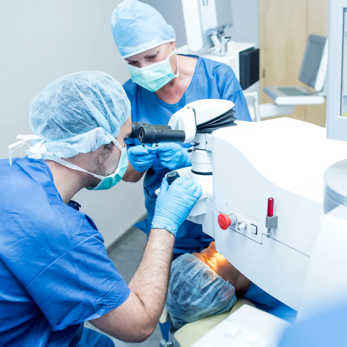
Preparation for an injection in a sterile environment

What is diabetic macular oedema?

Diabetic macular oedema is an accumulation of fluid at the centre of the retina, in the macula, in the diabetic patient. It occurs when vessels weakened by diabetes allow fluid to leak, which accumulates and thickens the retina, blurring fine vision. It is one of the leading causes of vision loss related to diabetes.

It can occur at different stages of diabetic retinopathy. The good news: effective treatments now exist, especially when the condition is managed early.

Diabetic macular oedema is a common complication of diabetes, but it is treatable. Regular screening allows it to be detected before any significant loss of vision.

Normal macula Thickened retina (oedema)
Schematic illustration inspired by an OCT cross-section: fluid thickens the macula. OCT allows this oedema to be measured and the response to treatment to be monitored.

Symptoms

Diabetic macular oedema mainly affects central vision:

  • blurred central vision;
  • distortion of straight lines;
  • difficulty reading or seeing fine detail;
  • colours appearing less vivid.

It can also be detected before any symptoms during screening, which underlines the importance of regular follow-up.

Diagnosis

OCT (optical coherence tomography) is the reference examination: it measures the thickness of the macula and the extent of the oedema precisely, without contact or discomfort, and allows the response to treatment to be monitored. This is where Dr Gattoussi's expertise in retinal imaging is particularly valuable.

  • OCT: measures and monitors the oedema;
  • Fundus examination: looks for signs of diabetes-related retinal damage (diabetic retinopathy);
  • Angiography: depending on the situation.

Treatments

Several treatments exist and may be combined. The choice depends on the extent of the oedema, the level of vision and the clinical context, and is discussed during the consultation.

Intravitreal injections

The most commonly proposed first-line treatment: a medication is injected into the eye to reduce the oedema, according to a monitoring protocol reassessed by OCT. See the page on intravitreal injections.

Laser treatment

Laser may be used in certain situations, alone or as a complement to injections.

Diabetes control

Essential and complementary to ocular treatments: controlling your diabetes and blood pressure, with your doctor, is a key factor in the outcome.

Management

Management combines treatment of the oedema and diabetes control, with close OCT monitoring.

  1. 1

    Diagnosis and OCT

    Measurement of the oedema and assessment of the associated retinopathy.

  2. 2

    Starting treatment

    Most often by intravitreal injections.

  3. 3

    Diabetes control

    In conjunction with your GP and diabetologist.

  4. 4

    OCT follow-up

    Regular reassessment to adapt the treatment frequency.

Learn more about intravitreal injections

Frequently asked questions

Can diabetic macular oedema be treated?

Treatments exist and are most effective when started early. Regular follow-up is essential.

Are injections repeated?

A monitoring protocol is put in place and adapted to the progress observed on OCT. The frequency may be higher at first, then reduced over time.

Is controlling diabetes alone sufficient?

It is essential, but often not sufficient on its own: it is combined with ocular treatments.

See also

Book an appointment

Consult Dr Gattoussi

Ophthalmology practice in Paris 11th. Online appointments via Doctolib, at any time. Sector 2 fees.