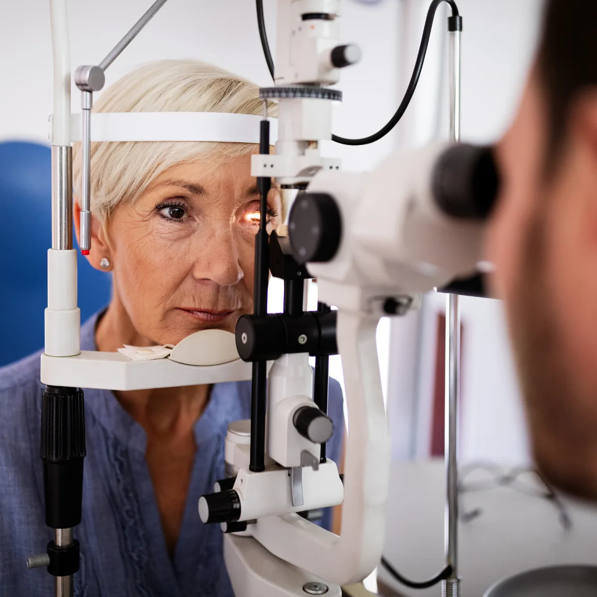
AMD is a disease of the macula, the central zone of the retina, which develops with age. It impairs fine vision, the vision used for reading and recognising faces, while peripheral vision is usually preserved. Regular monitoring allows the condition's progression to be followed.
AMD, or age-related macular degeneration, is a disease of the macula, the central zone of the retina responsible for fine and detailed vision. It develops with age, most often from the age of 55, and progressively impairs central vision, the vision used for reading, recognising faces, or driving. Peripheral vision is usually preserved.
AMD does not progress in the same way from one person to another. Regular screening and monitoring allow the progression to be followed and action taken at the right moment, particularly for the wet form.
AMD affects central vision, not peripheral vision: it does not lead to complete blindness, but can significantly impair reading and facial recognition.
Schematic illustration of the Amsler grid. In AMD, straight lines may appear wavy and a patch may appear at the centre. This self-monitoring tool does not replace an ophthalmic examination.
Forms of AMD
Two main forms are distinguished, which may coexist or follow one another. Distinguishing between them is essential, as their progression and management differ.
The dry form
The most common. It progresses slowly, over several years, through a gradual thinning of the macula. It does not require treatment by injections, but regular monitoring and preventive measures.
The wet form
Less common but potentially rapid in progression. It is linked to the appearance of abnormal blood vessels beneath the retina. It requires specific treatment, particularly intravitreal injections, and demands prompt management.
A sudden deterioration in vision or abrupt distortion of lines should prompt immediate consultation: it may indicate conversion to the wet form.
Causes and risk factors
AMD results from a combination of factors, some of which can be modified:
Age: the main factor, as the disease most often appears from the age of 55;
Family history: a hereditary predisposition is recognised;
Smoking: an important modifiable risk factor;
Lifestyle: diet, sun exposure, and cardiovascular risk factors are under study.
Symptoms
AMD can be silent, which is why screening is so important. Certain signs should prompt a consultation:
blurred central vision or a decline in reading vision;
distortion of straight lines (which appear wavy);
a dark patch or gap at the centre of vision;
a need for more light to read, or difficulty recognising faces.
The Amsler grid, illustrated above, is a simple self-monitoring tool between consultations. It does not replace an ophthalmic examination.
Diagnosis
Fundus examination and retinal imaging allow the diagnosis to be made, the form to be specified, and the progression to be monitored. This is where Dr Gattoussi's expertise in retinal imaging is particularly valuable.
OCT (optical coherence tomography): cross-sectional retinal imaging, painless and non-contact;
Fundus examination: examination of the macula;
Additional investigations (angiography, OCT-angiography) depending on the situation.
Management
Management depends on the form. The wet form is treated primarily by intravitreal injections, according to a regular monitoring protocol; the dry form is managed by surveillance and preventive measures. In all cases, follow-up is regular.
1
Diagnosis and imaging
Fundus examination and OCT to determine the form and activity of the disease.
2
Treatment decision
For the wet form, initiation of intravitreal injection treatment.
3
Treatment and reassessment
Injections are spaced out according to response, assessed by imaging.
4
Long-term monitoring
Regular follow-up of both eyes and self-monitoring with the Amsler grid.
Several lifestyle factors are under study in the prevention of AMD and its progression: stopping smoking, a diet rich in certain nutrients, protection from light, and control of cardiovascular risk factors. Dr Gattoussi discusses these during consultations.
AMD affects central vision but usually preserves peripheral vision: it does not lead to complete blindness. Monitoring aims to preserve vision for as long as possible.
Is AMD hereditary?
A family predisposition is recognised as a risk factor, alongside age and smoking. This is discussed during consultations.
What is the difference between the dry and wet forms?
The dry form progresses slowly and requires monitoring; the wet form can progress quickly and is treated with intravitreal injections.
Can AMD be prevented?
Age and heredity cannot be modified, but stopping smoking, a healthy lifestyle, and regular monitoring play an important role.