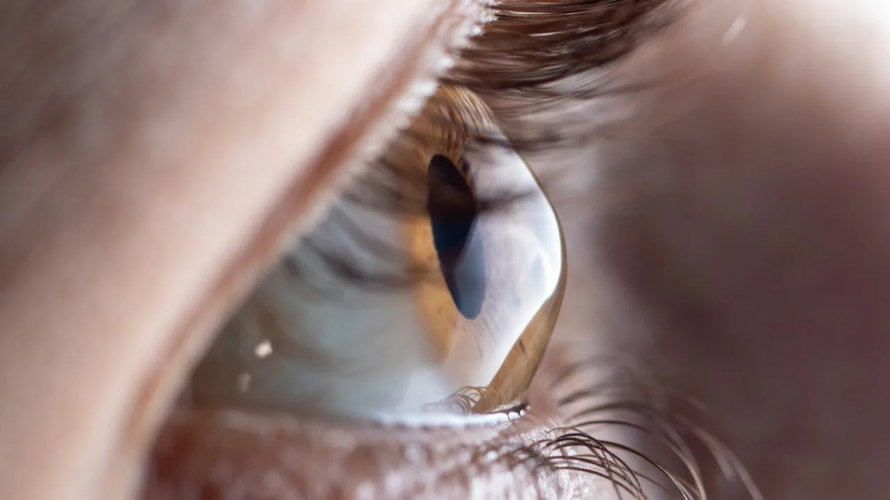
Keratoconus causes the cornea to thin and bulge into a cone shape. It often begins in adolescence and distorts vision. Detected early, it can be stabilised: that is the key challenge of screening.
In brief: keratoconus is a progressive deformation of the cornea, which thins and bulges. It causes progressive astigmatism and distorted vision. Eye rubbing aggravates it. Detected early by topography, it can be stabilised, and solutions exist to maintain good vision at every stage.
The cornea is the transparent lens at the front of the eye. Normally regular and round, it thins and deforms into a cone in keratoconus. This irregularity blurs and distorts images. The condition often begins in adolescence or in young adults and can progress over several years, at varying rates between the two eyes.
Frequent spectacle prescription changes in an adolescent or young adult should raise suspicion of keratoconus.
Keratoconus results from corneal fragility, to which several factors contribute:
The key examination is corneal topography, which maps the shape and thickness of the cornea and detects deformations even in their earliest stages, before they are otherwise visible. It is a painless, non-contact examination, particularly important in adolescents and before any planned refractive surgery. Dr Gattoussi's expertise in ocular imaging is especially valuable here.
Management has two objectives: stabilising the cornea and restoring good vision. It depends on the stage and rate of progression. Dr Gattoussi provides screening and follow-up of keratoconus, drawing in particular on corneal imaging; specialised treatments are carried out in dedicated facilities, to which she will refer you if needed.
By contrast, standard laser refractive surgery is generally contraindicated, as it would further weaken the cornea. See also: refractive surgery and pre-operative assessment.
Stopping eye rubbing is one of the most important measures to avoid aggravating keratoconus. In cases of allergy or itching, it is better to treat the underlying cause than to rub.
In the presence of increasing astigmatism, frequent prescription changes in a young person, or before any planned refractive surgery, a corneal assessment is recommended. The earlier keratoconus is detected, the greater the chance of stabilising it.
No. It can significantly impair vision, but does not cause blindness. Detected early, it can be stabilised, and solutions exist at every stage.
Yes, repeated rubbing is a recognised aggravating factor. Stopping eye rubbing is part of management.
Standard laser refractive surgery is generally contraindicated as it weakens the cornea. Other solutions are offered depending on the case.
A family predisposition is recognised: if there is a family history, screening of relatives, particularly adolescents, may be worthwhile.
This article is for general information purposes only and does not replace a consultation. It does not constitute individual medical advice.
Further reading